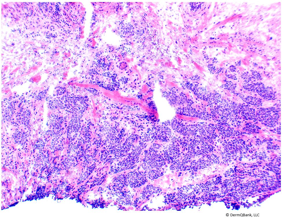
This is a Merkel cell carcinoma.
Merkel cell carcinoma (MCC), like atypical fibroxanthoma, is a rare rapidly
growing red nodule or plaque that is often ulcerated and favors chronically
sun-damaged skin of the head, neck, or legs in the elderly. They are rare tumors and considered to be fairly aggressive, with an overall 5-year survival (all stages) of ~60%. Treatment involves wide local excision and frequently
adjuvant radiation and chemotherapy.
(Coggshall et al. J Am Acad Dermatol 2018).
Histopathology of MCC shows poorly defined clusters, cords, and rosettes of small atypical mitotically
active blue cells throughout the dermis. The small atypical cells may demonstrate
single-cell necrosis and epidermotropism (the migration of malignant cells into
the epidermis). Other findings include a trabecular infiltrating pattern at the
periphery of the lesion, and crush artifact.
The staining of MCC is as follows:
- Positive: Neuron-specific enolase (NSE), epithelial membrane
antigen (EMA), synaptophysin, chromogranin
- Perinuclear dot staining pattern: Cytokeratin-20 (CK-20),
cam 5.2, AE-1, neurofilament
- Of note, CK-20 expression is almost entirely confined to the
gastric and intestinal epithelium, urothelium, and Merkel cells. CK-20
positivity is seen in colon adenocarcinomas, mucinous ovarian tumors,
transitional cell carcinoma, and Merkel-cell carcinoma.
- Negative: S-100, CEA, LCA
Cylindromas are uncommon smooth red nodules that are
typically seen on the scalp and can be associated with the Brooke-Spiegler syndrome. The histopathology of cylindromas shows a circumscribed
dermal nodule composed of a “jigsaw puzzle” arrangement of basaloid tumor
islands surrounded by a hyalinized eosinophilic PAS-positive basement membrane
cylinder. There are also hyalinized “droplets” and sweat duct lumina present
within the basaloid islands. The basaloid islands are composed of two
populations of cells, one with larger paler nuclei and the other with smaller
darker nuclei.
Basal cell carcinomas are classically a pearly red papule with overlying
telangiectasias. The classic histologic findings include: a basaloid tumor or strand budding
from the epidermis or hair follicles and/or within the dermis, retraction
artifact (stroma separation from basaloid nodules), peripheral palisading of
basaloid nuclei, stromal or basaloid lobules containing mucin, solar elastosis,
and variable inflammatory infiltrates. Basal cell carcinomas with multiple
nodules are arranged in a haphazard array rather than a tightly compacta
“jigsaw” pattern as is seen with a cylindromas.
Atypical fibroxanthoma (AFX) is an uncommon red nodule or plaque that appears on
chronically sun damaged skin of the head and neck in the elderly. It may be
considered a superficial variant of a malignant fibrous histiocytoma. Malignant
fibrous histiocytomas are deep subcutaneous or visceral masses clinically, as
opposed to the red ulcerated exophytic plaque or nodule resembling a nonmelanoma skin cancer seen
in AFX. The histopathology of AFX includes a dermal proliferation of
bizarre pleomorphic spindle cells, epithelioid cells, and multinucleated giant
cells closely abutting a thin epidermis. There is severe pleomorphism, multiple
typical and very atypical mitoses, and hyperchromatism.
Clinical Pearl: Merkel
cell carcinoma (MCC), is a rapidly growing aggressive tumor with high mortality
when metastasis occurs. Histopathology of MCC shows poorly defined clusters,
cords, and rosettes of small atypical mitotically active blue cells throughout the
dermis. Additional findings include: single-cell necrosis, epidermotropism, a
trabecular infiltrating pattern at the tumor periphery, and crush artifact.


